Office Closing - January 1st, 2024
Breast Imaging
What is a Mammogram?
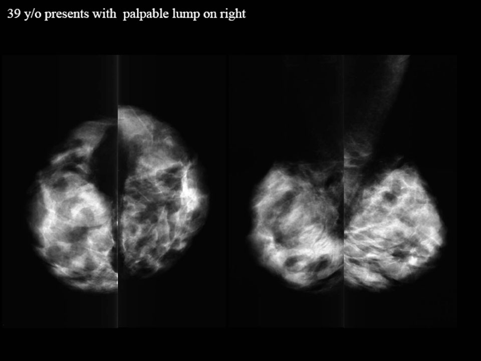
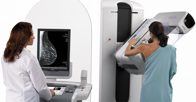
A mammogram is an X-ray of the breast. It is usually 2 views that are taken of each breast from different angles. Mammography is obtained by a special machine that uses a lower dose of radiation than the usual x-ray.
Mammograms are very good, but do have some limitations:
The compression of the breast that is required for good imaging can be uncomfortable
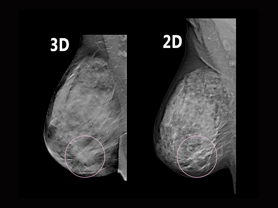
The compression also causes overlapping of the breast tissue. A breast cancer can be hidden in the overlapping tissue and not show up on mammogram.
Mammograms only take one picture across the entire breast in 2 directions.
Digital tomosynthesis is a special kind of mammogram that produces a 3-dimensional image of the breast. It takes multiple X-ray pictures of each breast from many angles. The breast is positioned in the same way as conventional mammogram, but only gentle pressure is applied. The X-ray tube moves in an arc around the breast will 11 images are taken during the 7 second exam.
What is a Breast Ultrasound?
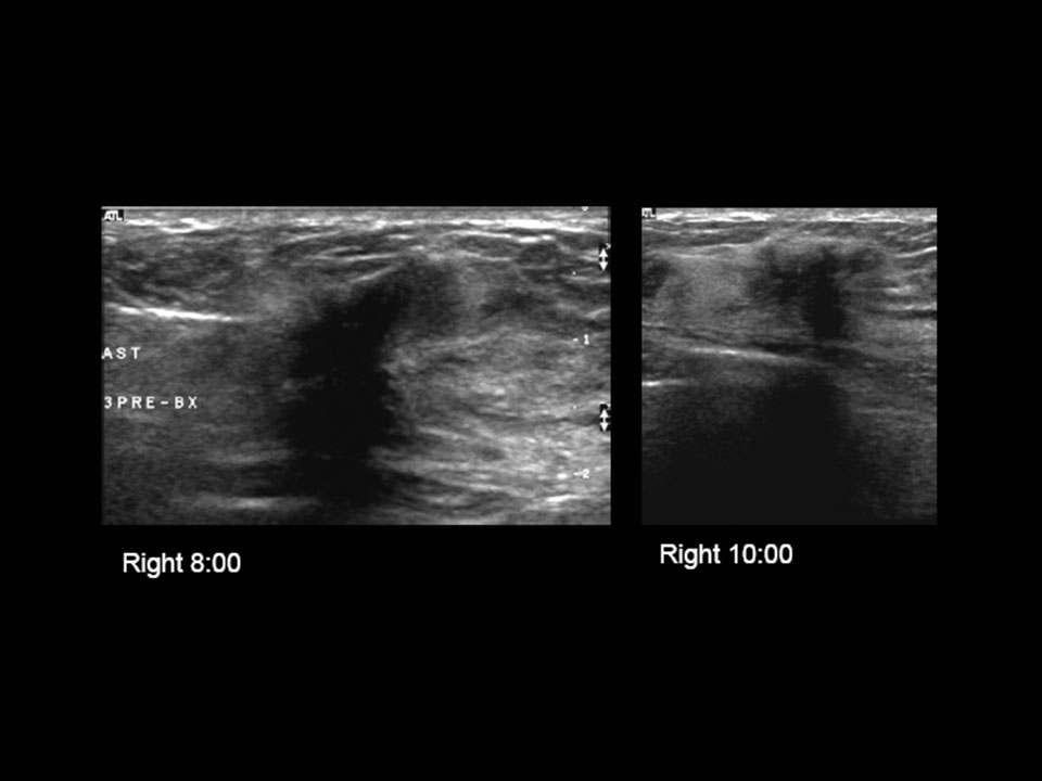
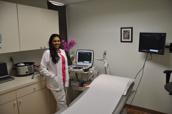
An ultrasound of the breast is a noninvasive and painless technique that creates two dimensional images used for the examination of the internal structures of the breast and the detection of abnormalities. With this, we can literally see the anatomy of the breast. Ultrasound utilizes a surface probe called a transducer that is held by the technician and requires no compression of the breast tissues as with mammography. The transducer emits sound wave energy into the breast which is then reflected back to the transducer creating a picture of the type and density of the tissues present. Ultrasound is used as an additional diagnostic tool when abnormalities are detected in the mammogram or physical examination. It is not intended as a replacement for mammography nor is it as cost effective as mammography as the initial screening tool due to the labor intensive nature of the imaging techniques. In the hands of a skilled sonographer with superior interpretive skills, the ultrasound can produce a highly reliable study. Since ultrasound does not utilize x-ray energy, it can be repeated more frequently and is completely safe for the patient.
What is a Breast MRI?
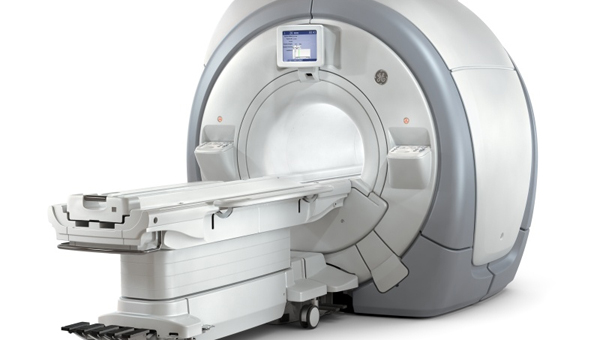
Magnetic resonance imaging (MRI) is an imaging tool that creates detailed, cross-sectional pictures of the inside of the body. Using radiofrequency waves, powerful magnets and a computer, MRI systems are able to distinguish between normal and diseased tissue.
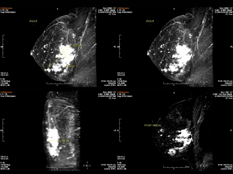
Breast MRI plays an important role in cancer diagnosis, staging and treatment planning. With MRI, we can distinguish between normal and diseased tissue to precisely pinpoint cancerous cells within the breast. It is also useful for revealing metastases. MRI provides greater contrast within the soft tissues of the body than a CT scan.
During an MRI, a patient rests on a table and slides into a large tunnel-shaped scanner. Breast exams usually require a contrast dye to be injected into a vein before the procedure. This helps certain areas show up better on the images. The procedure is painless and typically takes 30-60 minutes.
Unlike X-rays and CT scans, an MRI does not use radiation.
MRI for Breast Cancer
MRI technology for breast cancer uses radiofrequency waves to create detailed cross-sectional images of the breasts. MRI helps us to identify tumors in the breast that may have been missed by a mammogram.
